How 3D Dental Imaging Enhances Oral Health and Diagnostic Precision
November 19, 2025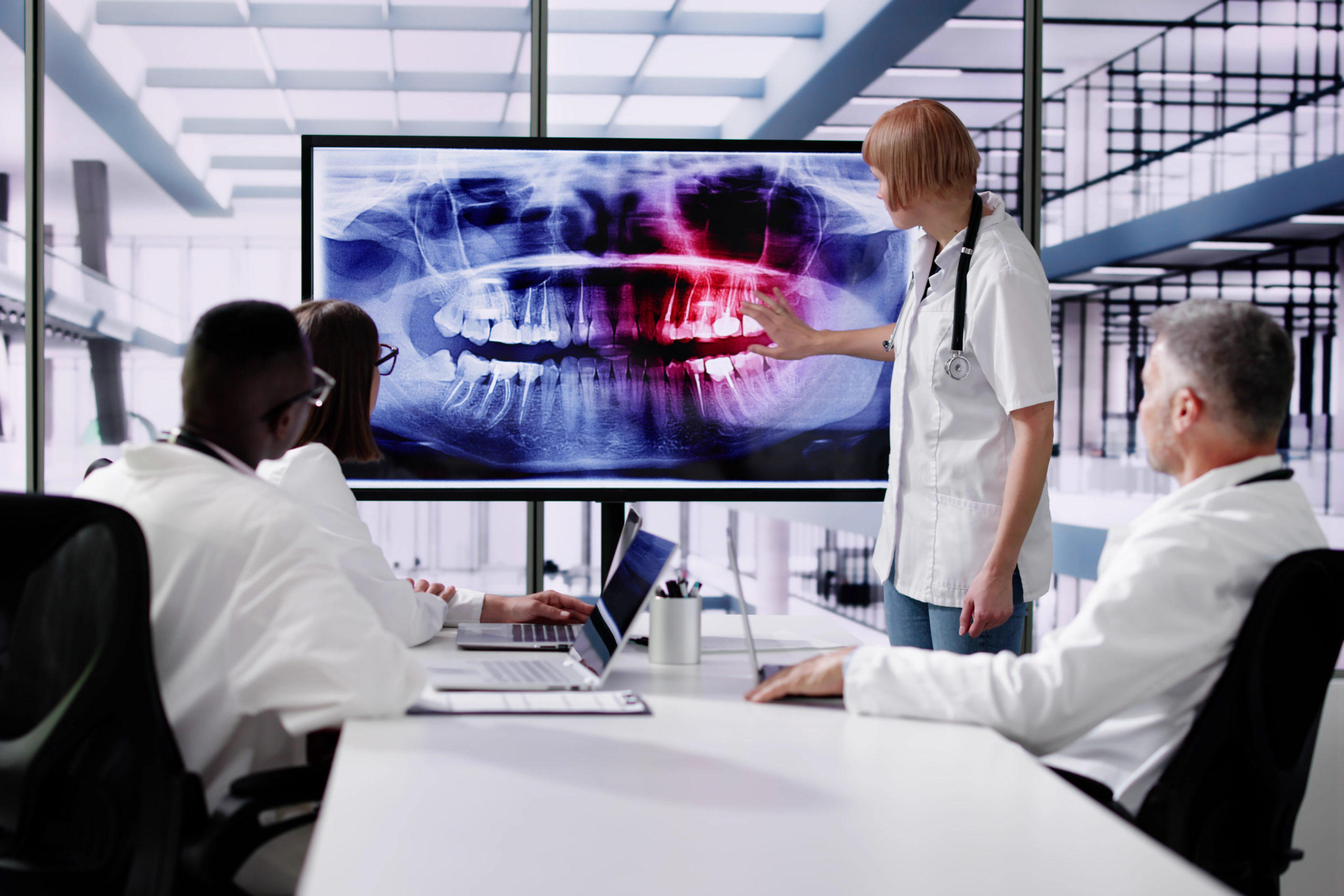
Technological advances have reshaped how dental professionals diagnose and treat oral health conditions. Dentists now rely on tools that provide granular insight, real-time accuracy, and more precise care. One of the most significant breakthroughs is 3D dental imaging, which is quickly becoming standard across clinics in the United States.
According to data from Fact.MR, the global market for 3D dental imaging is projected to hit $2.5 billion by 2024.
With the wide range of oral diseases and high patient expectations, clear and accurate diagnosis is non-negotiable in modern dentistry. 3D dental imaging changes the way professionals can detect problems, allowing for earlier intervention and better treatment results. With 3D dental imaging, dentists can get a detailed, three-dimensional view that traditional X-rays simply can’t provide, leading to smarter planning and greater confidence for both dental professionals and patients.
As adoption grows, more practices are learning the value of this technology for finding hidden issues and mapping personalized treatment. In this post, we’ll explain what 3D imaging is and how it stands apart from older imaging methods.
What Is 3D Dental Imaging?
3D dental imaging is one of the most transformative tools in dentistry today.
Unlike 2D X-rays, which show a single flat view, 3D imaging in dentistry creates detailed models of teeth, bone, nerves, and soft tissue. The technology most commonly used is called cone beam computed tomography (CBCT). CBCT is a specialized form of 3D radiography that provides layered, accurate, and complete images far beyond what is possible with older imaging.
With a 3D X-ray, the dental procedure involves rotating the scanner around the patient’s head, collecting hundreds of 2D images from different angles. Software then merges these into 3D dental images that capture even subtle issues within bone and tissue. The resulting 3D scan dental model offers a full view of oral anatomy, giving greater depth and clarity.
This shift to three-dimensional modeling helps dental professionals catch problems earlier and guides more accurate research and treatment. The level of depth and accuracy simply isn’t possible with traditional methods.
The Process: How 3D Imaging Works in Dentistry
Step 1 – Capturing the 3D Scan
The scanning process begins with a quick, comfortable session using a CBCT scanner.
The patient sits or stands in the unit, and the machine rotates around the head for less than a minute. During this time, it collects hundreds of individual 2D pictures from unique angles. These images build a precise 3D scan dental model. The process is painless, and the exposure to radiation is significantly lower than older full-mouth X-rays, reducing risk and most side effects.
Step 2 – Image Reconstruction and Visualization
Once the scan is complete, CAD/CAM software instantaneously compiles the data into high-resolution 3D dental images. Dentists can explore these models in real time, zooming in on any area of concern. We can view cross-sections, assess jaw joints, or analyze bone quality from every angle.
This depth of visualization means we catch issues like impacted teeth, fractures, or cysts not visible in a standard X-ray. This capability demonstrates how 3D imaging in dentistry improves the accuracy of every diagnosis.
Step 3 – Diagnosis and Treatment Planning
Dentists then use 3D radiography to guide every step of diagnosis and planning.
With more detailed imagery, professionals can determine the best way to place implants, identify hidden canals for root canal treatment, and plan orthodontic movement more precisely. The data also supports jaw analysis in complex surgery or trauma cases, such as evaluating the extent of mandibular fractures or planning corrective jaw (orthognathic) surgery.
Visual tools in the software let us share these findings with patients, so they can see the details first hand and better understand the recommended treatment.
Key Benefits of 3D Dental Imaging
The main benefit of 3D imaging in dentistry is the ability to diagnose problems with unmatched accuracy. Issues like hidden decay, early bone loss, root fractures, cysts, or tumors are far easier to spot. Guided implant surgery and custom restorations become safer, faster, and more reliable because we can plan every move based on a patient’s exact anatomy.
Real-time 3D X-ray dental imaging lets us make decisions without guesswork, especially for difficult procedures. Patients also get a clearer understanding of their condition through realistic digital visuals, leading to higher trust and compliance.
In the United States, clinics that adopt 3D dental imaging often see fewer complications and better long-term outcomes. The combination of precision, preventive value, and patient education is transforming modern oral health care.
Key benefits include:
- Enhanced detection of hidden dental issues such as decay, fractures, and bone loss
- Improved planning for implants, surgeries, and orthodontic treatments
- Real-time, detailed visuals for more accurate diagnoses
- Reduced guesswork during complex procedures
- Higher patient understanding and trust through clear digital imagery
- Fewer complications and better long-term outcomes
- Greater preventive care by identifying problems earlier
3D Dental Imaging vs. Traditional X-Rays
Comparing 3D dental imaging to conventional 2D X-rays reveals clear advantages for both dentists and patients. 3D imaging offers a full map of the mouth, fast diagnosis, and easy digital storage for later reference.
With traditional methods, images are limited to single angles and commonly miss hidden issues, making the process longer and less clear.
According to data from the National Institutes of Health, accuracy rates with 3D imaging are around 90% overall, compared to around 70% with 2D imaging. Here are the most prominent differences:
| Feature | 2D Imaging (Conventional X-Rays) | 3D Imaging (CBCT/3D X-ray) |
| Dimensionality | Flat (two-dimensional) | Volumetric (three-dimensional) |
| Detail Level | Limited, may obscure overlapping structures | Reveals subtle details, no overlap of anatomy |
| Visualization | Only specific slices or angles | Rotatable, full view of oral anatomy |
| Diagnostic Value | Good for general screening | Superior for complex cases (e.g., periodontitis) |
| Radiation Dose | Generally lower | Slightly higher, but justified for complex cases |
| Application | Routine exams | Implant planning, pathology, periodontitis |
Modern 3D radiography uses much less radiation than past panoramic X-rays. Newer CBCT machines have settings that keep doses as low as possible, adding to patient peace of mind. These advantages explain why many dental clinics throughout the United States are moving to 3D systems for daily care.
Applications of 3D Imaging in Dentistry
3D imaging has revolutionized many areas of dental practice by offering precise and comprehensive views of oral structures.
Implant Dentistry
3D imaging delivers more accurate models for implant placement for better safety and proper bone density assessment.
By visualizing anatomical structures in three dimensions, clinicians can identify optimal implant sites and avoid critical areas such as nerves and sinuses. This improves patient outcomes by reducing surgical complications, shortening procedure times, and increasing the predictability of implant integration with the surrounding bone.
Orthodontics
For orthodontic treatment, full-jaw 3D scans show detailed root positions and jaw relationships, resulting in more precise treatment planning and alignment.
These scans help identify impacted or missing teeth, evaluate airway space, and assess bite discrepancies from multiple angles. The enhanced visualization helps with the customization of orthodontic appliances and allows for the monitoring of treatment progress with a high degree of accuracy, ultimately leading to better long-term results.
Endodontics
Endodontic specialists use 3D dental images to detect accessory canals, root fractures, and other complications that may affect root canal procedures.
This technology allows for a comprehensive assessment of the internal anatomy of teeth, which is often missed on traditional 2D radiographs. By identifying hidden infections, calcifications, or unusual curvatures, clinicians can plan more effective and less invasive treatments, improving the success rate of endodontic therapy.
Oral Surgery
Oral surgeons depend on 3D scan data to plan safe tooth extractions and reconstructive surgeries with greater accuracy.
The ability to visualize the exact position of impacted teeth, bone defects, and adjacent anatomical structures like nerves or blood vessels can work to reduce intraoperative risks. Detailed preoperative planning also helps optimize graft placement and predict surgical outcomes, contributing to quicker recovery and minimized patient discomfort.
Dental Prosthetics
The advancement of 3D printing for dental crowns and restorations relies on detailed scan models, allowing for a precise fit and better results. With 3D imaging, dental labs can fabricate prosthetics that closely match the patient’s natural dentition and bite dynamics.
This technology streamlines the workflow from impression to final placement, reduces the need for manual adjustments, and increases patient satisfaction with both the appearance and function of their dental prostheses.
The Future of 3D Imaging in Oral Health
The future of oral diagnostics is heading toward smarter, faster, and more precise results in dental health. AI-assisted analysis of 3D scans will soon help catch issues even earlier. Direct integration with CAD/CAM design software will automate parts of the process and save significant clinical time.
At the same time, further improvements in 3D radiography are shrinking radiation exposure and increasing sharpness. As digital dentistry keeps advancing, we’ll continue to see better results in both preventive and restorative dental care.
Frequently Asked Questions About 3D Dental Imaging
Is 3D dental imaging safe for patients?
Yes, 3D dental imaging with CBCT uses low, safe levels of radiation compared to many traditional imaging methods. The technology is designed to minimize unnecessary exposure, and protocols are in place to ensure that scans are only performed when clinically justified. Patients can feel confident in the safety of this diagnostic tool, which balances the need for detailed images with radiation protection standards.
How long does a 3D dental scan take?
The actual scan usually takes less than a minute, and the whole process (setup to review) rarely exceeds ten minutes. Most of this time is spent on patient positioning and post-processing of images. The efficiency of 3D scanning means minimal time in the dental chair, allowing for a comfortable experience and quick results that can be discussed with your dentist during the same visit.
Does a 3D dental X-ray expose me to more radiation?
No, most modern 3D scans use equal or lower radiation levels than traditional full-mouth X-rays. The equipment is specifically calibrated to deliver only the necessary dose for diagnostic clarity. Advances in CBCT technology have further reduced radiation exposure, adhering to the ALARA (As Low As Reasonably Achievable) principle and making it a safe choice for most dental patients.
When is a 3D scan necessary in dentistry?
3D scans are valuable for dental implants, complex root canals, orthodontic evaluation, and planning any surgical procedure. They provide critical information that cannot be obtained from 2D imaging alone. Dentists may also recommend 3D scans for the assessment of jawbone health, detection of cysts or tumors, and evaluation of impacted teeth, ensuring comprehensive treatment planning and precise execution.
Can 3D dental imaging detect all oral health problems?
While 3D imaging shows far more detail than 2D X-rays, some conditions still require other tests or clinical exams for full diagnosis. For example, soft tissue lesions or early-stage decay may not be visible on CBCT scans. Dentists typically use 3D imaging as part of a broader diagnostic approach, combining it with traditional exams and additional tests to ensure nothing is missed.
Want to Learn More?
In modern dental practices, 3D dental imaging is a powerful tool for improving diagnostic accuracy, treatment, and patient trust. The level of detailed insight provided by this technology leads to more predictable outcomes and less risk during complex procedures.
Adopting 3D imaging is a step toward higher standards of preventive and restorative care: an advantage that will only continue to grow.
Experience the precision of advanced 3D imaging. Schedule a consultation with Cad-Ray to see how innovation is shaping healthier smiles.